Plant Cell Under Microscope : Plant Cell Under The Microscope View Stock Image Image Of Macro Biological 153600001 : Image:plant cell seen under electron microscope.
byJonah Laycock-
0
Plant Cell Under Microscope : Plant Cell Under The Microscope View Stock Image Image Of Macro Biological 153600001 : Image:plant cell seen under electron microscope.. Your plant cells under microscope stock images are ready. Plant cell structure under microscope : Microscopic photography plant cell cell biology microbiology science art organic shapes botany 2d design textiles. A cell is a very tiny structure which exists in living bodies. In this premade slide of vicia pea's root, you can see the active cell division at the tip of a growing root.
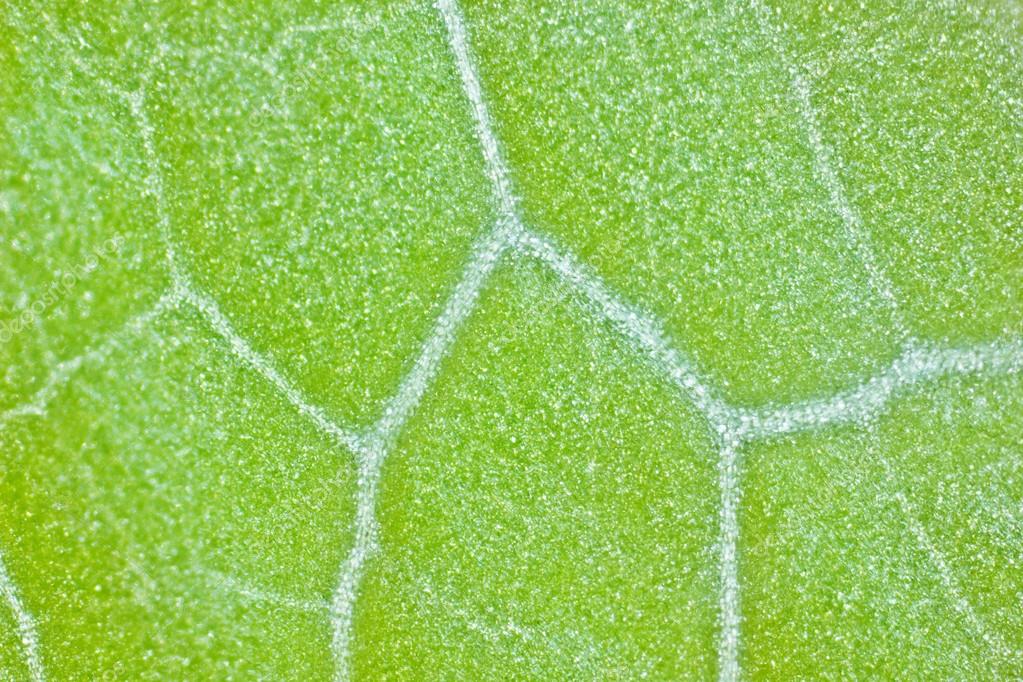
Plant cell structure under microscope : Under a x400 light microscope we could see the cell wall, cell membrane, nucleus and cytoplasm the parts of a (palisade) plant cell that can be seen under a light microscope are:cell wallcell (surface) membranelarge (permanent) vacuolecytoplasmnucleuschloroplasts. The growth and development of the cork cambium tissue vary with the plant species in consideration. Chloroplast flowing inside the plant cell under 100x len. To learn how to get the best image from a microscope.
Plant Cells Under Microscope Under Microscope Cells Under Microscope Plant Cell from i.pinimg.com The high resolving power makes the electron microscope a very important research tool in microbiology. Who doesn't like seeing cool stuff like human skin cells, dog hair and pond scum magnified before their eyes? Their distinctive features include primary cell walls containing cellulose, hemicelluloses and pectin, the presence of plastids with the capability to perform photosynthesis and store starch. Tulip stem cells at the. Select the lowest power objective lens. It also depends on plant age and conditions of plant growth. The growth and development of the cork cambium tissue vary with the plant species in consideration. Chloroplast cells under a microscope | educational fun.
Examining plant cells under the microscope.
Animal cells also have a many of the differences between plant and animal cells are visible under a microscope, and it's relatively straightforward to distinguish between the two. Who doesn't like seeing cool stuff like human skin cells, dog hair and pond scum magnified before their eyes? Their distinctive features include primary cell walls containing cellulose, hemicelluloses and pectin, the presence of plastids with the capability to perform photosynthesis and store starch. The growth and development of the cork cambium tissue vary with the plant species in consideration. In this simple microscope experiment, we will compare plant cells and animal cells. 9 pupil activity cell structure read through the information on each of the organelles as you colour them in follow the guidance on colouring them in given at the bottom of the page this works on the theory that whilst you are. Plant cells under the microscope. Find the perfect plant cells under microscope stock photos and editorial news pictures from getty images. Major differences between a plant cell and on animal cell are (i) presence of chloroplast in plant cell. Guide to plant anatomy and biology using microscope premade slide sets. Image:plant cell seen under electron microscope. Observe the onion skin under low power of the microscope and then under high power. My students used to love microscope lab days.
Use them in commercial designs under lifetime, perpetual & worldwide rights. Under a x400 light microscope we could see the cell wall, cell membrane, nucleus and cytoplasm the parts of a (palisade) plant cell that can be seen under a light microscope are:cell wallcell (surface) membranelarge (permanent) vacuolecytoplasmnucleuschloroplasts. Plant cells are eukaryotic cells present in green plants, photosynthetic eukaryotes of the kingdom plantae. Chloroplast flowing inside the plant cell under 100x len. To learn how to get the best image from a microscope.
Plant Cells Under Microscope Stock Photo By C Solstudio 103192844 from st2.depositphotos.com Resolving power is the ability to distinguish between separate things which are close to each other. Generalized cell is used for structure of animal cell and plant cell to present the common parts, appearing in various parts of the bodies of animals and plants. Place the glass slide onto the stage. Use them in commercial designs under lifetime, perpetual & worldwide rights. It also has a very high resolving power. Guide to plant anatomy and biology using microscope premade slide sets. The high resolving power makes the electron microscope a very important research tool in microbiology. Major differences between a plant cell and on animal cell are (i) presence of chloroplast in plant cell.
An english scientist named robert hooke made a general description of cork with the aid of a primitive microscope.
In contrast to normal cells, cancer cells often exhibit much more variability in cell size— some are larger than normal and some are smaller than. Pink plant cells under microscope. Examining plant cells under the microscope. It also has a very high resolving power. (ii) presence of large central vacuole in plant cell. Their distinctive features include primary cell walls containing cellulose, hemicelluloses and pectin, the presence of plastids with the capability to perform photosynthesis and store starch. Plant cell structure under microscope : 9 pupil activity cell structure read through the information on each of the organelles as you colour them in follow the guidance on colouring them in given at the bottom of the page this works on the theory that whilst you are. Plant cells are eukaryotic cells present in green plants, photosynthetic eukaryotes of the kingdom plantae. 8 pictures of plant cells under a microscope. Onion epidermis under light microscope. Chloroplast cells under a microscope | educational fun. To look at a cell close up we need a microscope.
Select the lowest power objective lens. 50 amazing things under electron microscope sem images in this video you can see 50 amazing that are seen and captured. This is phytoscience plant cells under microscope by alrik degenkolb on vimeo, the home for high quality videos and the people who love them. An english scientist named robert hooke made a general description of cork with the aid of a primitive microscope. See more ideas about microscopic photography, plant cell, microscopic.
Animal Cell Under Microscope Structure And Anatomy from www.thegreatestgarden.com Animal cells also have a many of the differences between plant and animal cells are visible under a microscope, and it's relatively straightforward to distinguish between the two. Plant cells are eukaryotic cells present in green plants, photosynthetic eukaryotes of the kingdom plantae. Appearance —under a microscope, normal cells and cancer cells may look quite different. Plant cell organelles that are invisible under a compound light microscope include mitochondria, ribosomes, endoplasmic reticula, and golgi bodies. Find the perfect plant cells under microscope stock photos and editorial news pictures from getty images. Your plant cells under microscope stock images are ready. Here's a photo of a plant cell under an electron microscope. Place the glass slide onto the stage.
(iii) presence of cell wall.
Lets get looking at some real plant cells! Appearance —under a microscope, normal cells and cancer cells may look quite different. The growth and development of the cork cambium tissue vary with the plant species in consideration. (iii) presence of cell wall. Plant cells under the microscope. Your plant cells under microscope stock images are ready. Find the perfect plant cells under microscope stock photos and editorial news pictures from getty images. 8 pictures of plant cells under a microscope. To look at a cell close up we need a microscope. Under high magnification, you can even identify cells undergoing mitosis, and different phases of mitosis. Who doesn't like seeing cool stuff like human skin cells, dog hair and pond scum magnified before their eyes? It also has a very high resolving power. Image:plant cell seen under electron microscope.